Ball and Socket Mechanism
To hold the probe and allow probe insonation angles to be adjusted, we chose a ball and socket mechanism with a threaded hole along the diameter of the ball. The hole on the ball is threaded inside, so the probe, which has a counter-threaded body, can be inserted into the ball. The threads allow the probe to be inserted and stopped at any position along the hole as needed. The ball, with the probe inserted, is allowed to rotate in the socket with three degrees of freedom in a spherical coordinate system. The bottom of the ball and socket are cut flat so the probe, ball and socket can all lay flush to the patient’s head. The height of the socket is kept at a minimum to generate a wider opening on the top of the socket in order to allow wider rotation. However, it is not so wide that the ball falls out.

Ball and Socket-Plate Body 3D
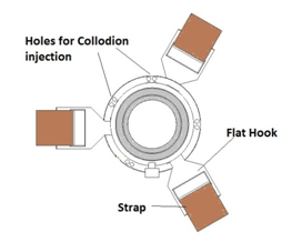
To hold the probe steadily against the patient’s temporal window, both elastic straps (mechanical) and adhesives (chemical) are adopted into the system to secure the pedestal on patient’s head. The bottom of the pedestal has a circular plate with a hole at the center, allowing the probe to access the patient’s head. The elastic straps are the primary fixing mechanism to secure the pedestal in place. The three straps, with flat hooks at each end, hook onto rectangular indents at the edge of the circular plate, located 120 degrees apart.
In addition to the straps, collodion is used as a secondary fixation that prevents minor dislocation of the pedestal during pedestal installation and the course of the therapy. Collodion, a solution of pyroxylin, ether, and alcohol, can be applied to the twelve holes on the plate, gluing the pedestal to the skin. The twelve holes are grouped into four sets and are located as shown below to maximize stability.
In addition to the straps, collodion is used as a secondary fixation that prevents minor dislocation of the pedestal during pedestal installation and the course of the therapy. Collodion, a solution of pyroxylin, ether, and alcohol, can be applied to the twelve holes on the plate, gluing the pedestal to the skin. The twelve holes are grouped into four sets and are located as shown below to maximize stability.
Top view of the Plate
Each pedestal assembly is made of a ball, a socket-plate body with a thumbscrew, and three straps with hooks (6 hooks). To form an entire system, two pedestals are used on each side of patient’s head right above his/her temporal window.

Schematic of Plate and Socket
